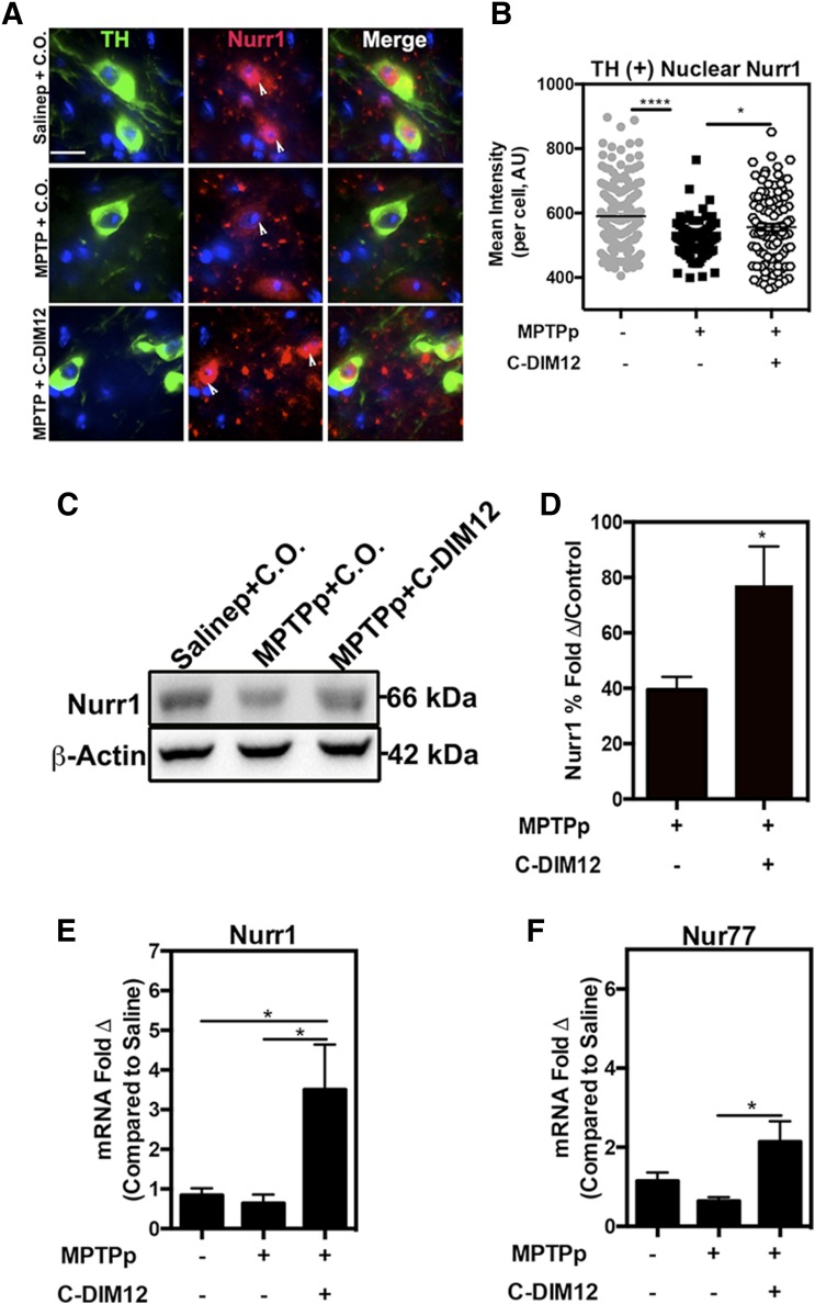
Fig. 7.
Subcellular localization and expression of Nurr1 are modified by C-DIM12 treatment in vivo. (A) The 100× objective images of TH+ neurons (green) show that Nurr1 (red) is sequestered to the nucleus with C-DIM12 treatment, and white arrows depict nuclear localization. (B) Mean intensity of TH+ nuclear Nurr1 is significantly higher in the C-DIM12 group compared with the MPTP + C.O. group (*P < 0.05; ****P < 0.0001; N = 4 animals/group). Western blot of Nurr1 protein isolated from ST tissue shows that C-DIM12 prevents MPTPp-induced protein changes (C), as illustrated in the quantitative measurement of mean optical density (D) (control set to 100%; *P < 0.05; N = 6–8 animals/group). qPCR data of mRNA isolated from SNpc for Nurr1 (E) and Nur77 (NR4A1) (F) expression show C-DIM12 induces higher levels of NR4A2 (*P = 0.05; N = 8 animals/group).

