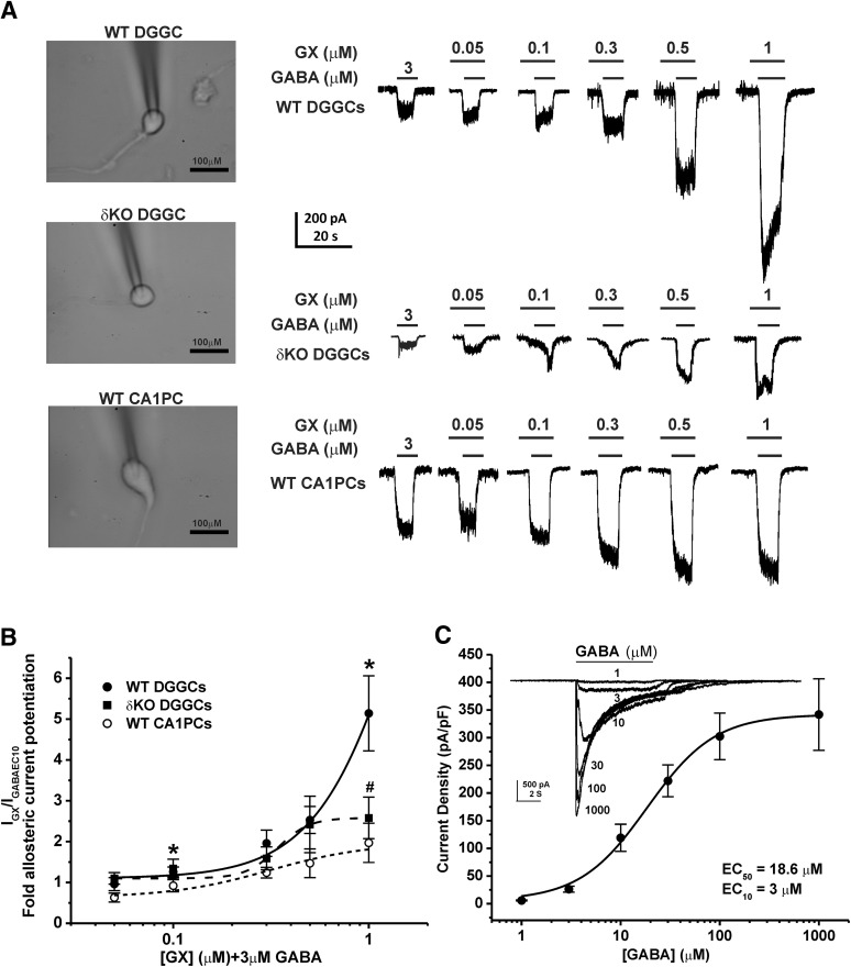
Fig. 2.
GX allosteric activation of GABA-gated currents in acutely dissociated neurons. GX (1 μM) displayed significantly greater GABA-gated chloride currents in WT DGGCs (5.1 ± 0.9-fold potentiation) than CA1PCs (2.0 ± 0.5-fold potentiation). However, GX-potentiated GABAergic currents were significantly reduced in δKO DGGCs. (A) Representative whole-cell current recordings of DGGCs and CA1PCs. Neurons displayed concentration-dependent responses to GX potentiation of 3 μM GABA (EC10). (B) Concentration-response of GX-modulated allosteric potentiation of chloride currents in DGGCs and CA1PCs from wild-type or δ-subunit knockout (Gabrd−/−, δΚΟ) mice. (C) Concentration-response profile of GABA-potentiated current density. GABA EC10 = 3 μM. Each point represents mean ± S.E.M. of data from 5–10 cells. *P < 0.05 vs. CA1PCs; #P < 0.05 vs. WT DGGCs.

