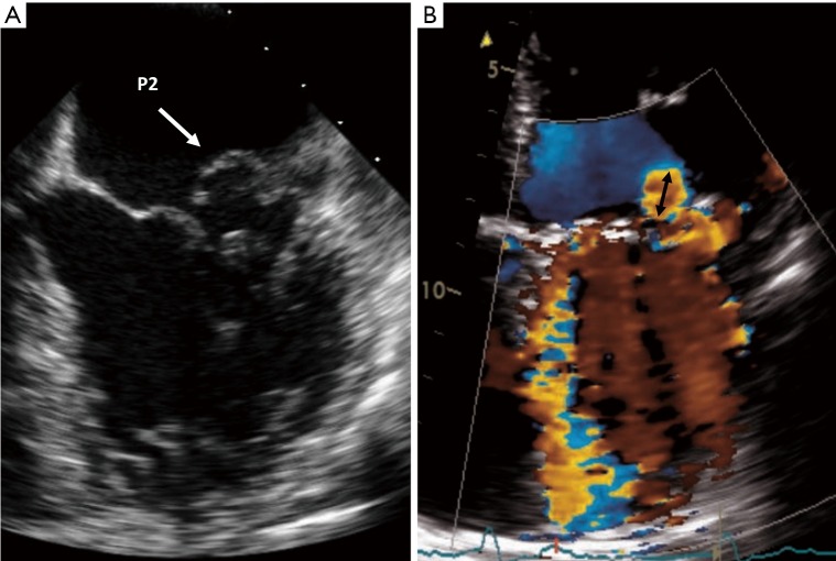
Figure 2.
Echocardiography demonstrating mitral valve prolapse and its associated color Doppler. (A) Mitral valve posterior leaflet (P2) prolapse seen in transesophageal echocardiogram. (B) Eccentric, wall-impinging jet of MR with Coanda effect. Although the jet area is small, but the PISA radius (black arrow) is large and alert to the severity of regurgitation.

