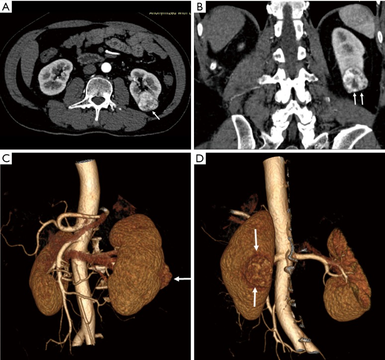
Figure 1.
Contrast-enhanced CT images with 2D and 3D reconstructions showing malignant renal tumour. (A) 2D axial CT image shows a tumour with contrast enhancement at the lower and posterior region of left kidney (arrow); (B) coronal reformatted view shows the tumour’s enhancement is heterogeneous (arrows), with low-attenuation areas within the lesion; (C) 3D volume rendering frontal view shows the tumour is located at the posterior aspect of left kidney (arrow); (D) 3D volume rendering posterior view shows the tumour location (arrows). CT, computed tomography; 2D, two-dimensional; 3D, three-dimensional.

