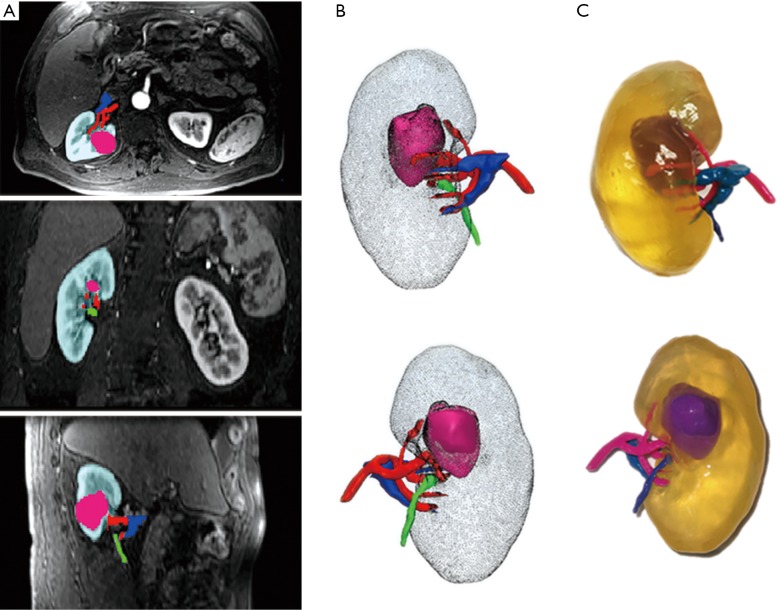
Figure 3.
Use of contrast-enhanced MR images for generating 3D printed model of renal tumour. (A) Axial, coronal, and sagittal views of MRI images with segmentation masks for one representative case. Kidney = teal, tumour = pink, artery = red, vein = blue, collecting system = green; (B) anterior and posterior 3D projections. Kidney = gray, tumour = pink, artery = red, vein = blue, ureter = green; (C) photographs of 3D printed model. Kidney = transparent, tumour = purple, artery = pink, vein = light blue, ureter = dark blue. Reprinted with permission from Wake et al. (18). MRI, magnetic resonance imaging; 3D, three-dimensional.

