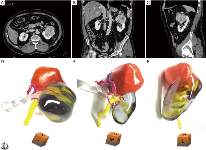
Figure 4.
3D printed model for case 1 who is a 67-year-old male with renal tumour at the upper pole of left kidney. Comparative views of the CT scan at the nephrographic phase [(A) axial, (B) coronal and (C) sagittal planes] and corresponding views of the physical model [(D) superior and median view, (E) median and anterior view, (F) lateral view]. An inferior polar cyst is also displayed on this model (translucent yellow). The cubes show the 3D printed model orientation in space (I = inferior face, A = anterior face, L = lateral side, S = superior face, P = posterior face, M = median side). Case 1 underwent a left radical nephrectomy for a 65×56×42 mm clear cell renal cell carcinoma, pT1bN0Mx, Fuhrman grade 3. The arterial tree is presented in opaque magenta, the collecting system in opaque yellow, and opaque orange for tumour display. The renal vein and renal parenchyma are kept translucent to allow the best visualization of the relationships between the renal tumour and surrounding structures. Reprinted with permission from Bernhard et al. (15). 3D, three-dimensional.

