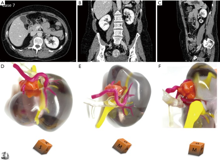
Figure 5.
3D printed model for case 7 who is a 53-year-old female with renal tumour at the interpolar region of left kidney. Comparative views of the CT scan at the nephrographic phase [(A) axial, (B) coronal and c sagittal planes] and corresponding views of the physical model [(D) superior view, (E) median view, (F) median view]. The cubes show the 3D printed model orientation in space (I = inferior face, A = anterior face, L = lateral side, S = superior face, P = posterior face, M = median side). Case 7 underwent a left partial nephrectomy for a 21×15×15 mm angiomyolipoma. Description of colour corresponding to different renal structures and tumour is the same as in Figure 4. Reprinted with permission from Bernhard et al. (15). CT, computed tomography.

