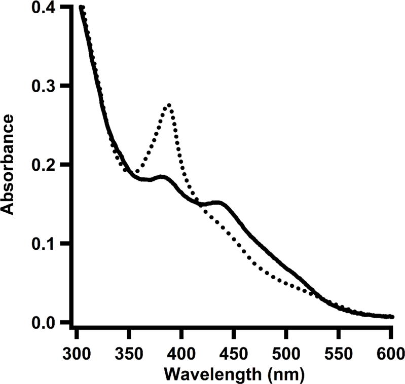
Figure 4.

UV-vis spectrum of TsrM treated with dithionite. Reduced form of enzyme (dotted line) shows a predominant peak at 390 nm, corresponding to cob(I)alamin. Upon addition of SAM (solid line), the cob(I)alamin peak decreases and a new peak arises at 450 nm, which corresponds to the formation of methylcobalamin in its base-off/His-off form.
