
Fig. 5.
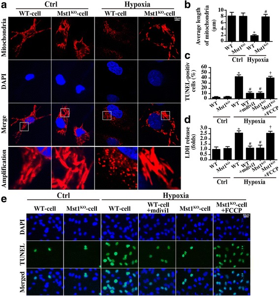
Mst1 controlled cellular apoptosis via mitochondrial fission. a The mitochondria of wild-type cells (WT-cell) and Mst1-knockout cells (Mst1KO-cell) were stained with Tom20 under control (Ctrl) and hypoxia conditions and mitochondrial fission was measured. b The average length of mitochondria was quantified. c and e The TUNEL assay was used to label the apoptotic cells. d The LDH release assay was carried out to measure the cardiomyocyte damage with mitochondrial fission activation and inhibition. *p < 0.05 vs. ctrl group, #p < 0.05 vs. WT-cell+hypoxia, †p < 0.05 vs. Mst1KO-cell+hypoxia