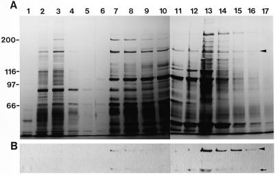
Figure 2.
Hydroxylapatite column chromatography of the ATP extract. A, Silver staining of a 6% (w/v) SDS-polyacrylamide gel. B, Immunoblotting of the same fractions using AS-170. Numbers on the top of gels indicate fraction numbers. After the application of the ATP extract shown in Figure 1B on a hydroxylapatite column, the adsorbed materials were eluted with a discontinuous gradient of 5 mm (fraction 1–5), 150 mm (fraction 6–11), and 300 mm (fraction 12–17) potassium phosphate buffer. The arrowheads and arrow indicate 170-kD myosin and the 120-kD degradation product of the 170-kD heavy chain, respectively.

