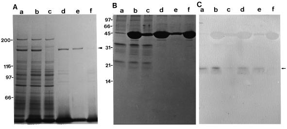
Figure 6.
Co-precipitation experiment of polypeptides in DE-52 column fractions with F-actin. A, Silver staining of a 6% (w/v) SDS-polyacrylamide gel. B, Silver staining of a 15% (w/v) SDS-polyacrylamide gel. C, Immunoblotting of the same samples shown in B with AS-CaM. Fractions 9 to 15 shown in Figure 4 were pooled (lanes a) and mixed with F-actin (lanes b). Supernatant (lanes c) and pellet (lanes d) after centrifugation of the mixture shown in lanes b. Supernatant (lanes e) and pellet (lanes f) after ATP treatment of the pellet shown in lanes d. The arrowhead and arrow indicate the 175-kD polypeptide and the 18-kD band, respectively. The percentage of translocated F-actin in motile assay was 79%, 2%, and 92% for fractions a, c, and e, respectively.

