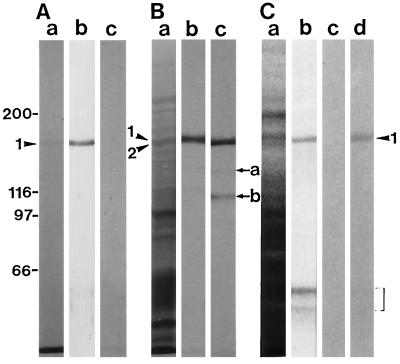
Figure 8.
Cross-reactivity of antibodies against the 175-kD myosin heavy chain. A, Immunoblotting of isolated 175-kD myosin shown in Figure 6. Lane a, Coomassie Brilliant Blue staining of a 6% (w/v) SDS-polyacrylamide gel; lane b, immunoblotting using AS-175; lane c, immunoblotting using AS-170. B, Immunoblotting of the ATP extract of the co-precipitant with F-actin in a crude cell extract. Lane a, Silver staining of a 6% (w/v) SDS-polyacrylamide gel; lane b, immunoblotting using AS-175; lane c, immunoblotting using AS-170. C, Immunoblotting of a crude protein sample from BY-2 cells. Lane a, Coomassie Brilliant Blue staining of a 6% (w/v) SDS-polyacrylamide gel; lane b, immunoblotting using AS-175; lane c, immunoblotting using non-immune serum; lane d, immunoblotting using AAB-175. The arrowheads 1 and 2 indicate the 175- and the 170-kD myosin heavy chains, respectively. The arrows a and b indicate the 135- and the 120-kD degradation product of the 170-kD myosin heavy chain, respectively. The region marked by a bracket shows the low-molecular-mass polypeptides reacting with AS-175.

