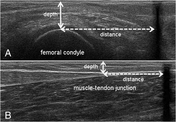
Fig. 2.

Ultrasound images used to assess the GM muscle belly and Achilles tendon lengths, based on the US-tape method of Barber et al. 2011. The dashed lines show the distance from the superficial aspect of the condyle (a) and the most distal aspect of the muscle-tendon junction (b) to the tape (black shadow), whereby the white arrows indicate the US depth
