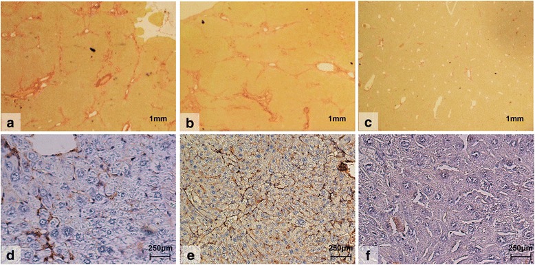
Fig. 3.

Histological images using Picro-sirius red staining (a-c) of liver tissue for fibrosis and immunohistochemical staining for αSMA (d-f) in mice treated with 8 weeks of thioacetamide (TAA) with or without FXa or thrombin inhibition. Control mice treated with TAA (n = 13) alone (A; × 15magnification). Mice treated with TAA and the thrombin inhibitor (n = 12), Dabigatran (100 mg/kg) (B; × 15 magnification). Mice treated with TAA and the FXa inhibitor (n = 13), Rivaroxaban (40 mg/kg) (C; × 15 magnification)
