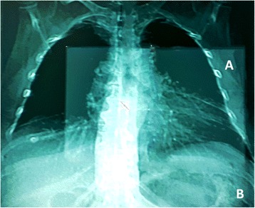
Fig. 1.

Ultrafast single protocol. The ECG-gated helical prospective acquisition started from the carena to the apex of the heart to evaluate coronary arteries (a, field of vew of cardiac scan), followed by fast, low dose acquisition, from pulmonary apex to the bases, on the whole chest (b, field of view of thoracic scan)
