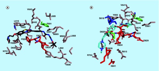
Figure 8. . FLT3 residues that are near the bound ligands.
(A) FLT3 residues that are within 6 Å of quizartinib in the docked structure; (B) FLT3 residues that are within 6 Å of HSN286 in the docked structure. The residues on the A loop including the DFG motif are shown as red while the P loop residues are colored blue.
FLT3: FMS-like tyrosine kinase 3.

