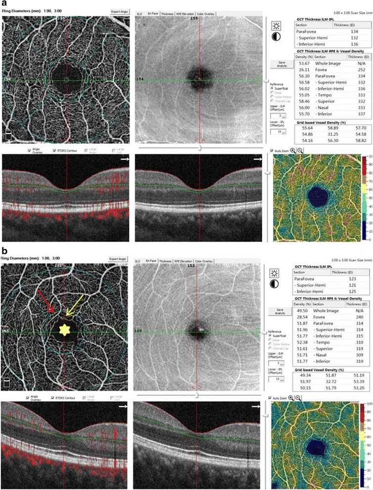
Fig. 1.

OCTA images of retinal structures and vessel networks, with reports of retinal thickness and vessel density, in a normal subject and a diabetic patient (3 × 3 mm scan area). a The structural OCT and angiography of a normal subject did not show abnormalities and the image quality was good. b The structural OCT of a diabetic patient did not show abnormalities. Capillary loss (yellow arrow), morphological anomalies (red arrow), and deformed foveal avascular area (yellow star) were demonstrated in an angiography scan
