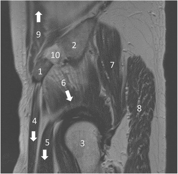
Fig. 4.

MRI of a pelvis and the hip joint in T2 sagittal reconstruction: 1 = ASIS, 2 = iliac crest, 3 = femoral head, 4 = sartorius muscle, 5 = femoral rectus muscle, 6–8 = minor, medium, and greater gluteal muscles, 9 = abdominal muscle, 10 = location of bone graft harvesting. The arrows show the traction of muscle forces that provoke a fatigue fracture of the ASIS [13]
