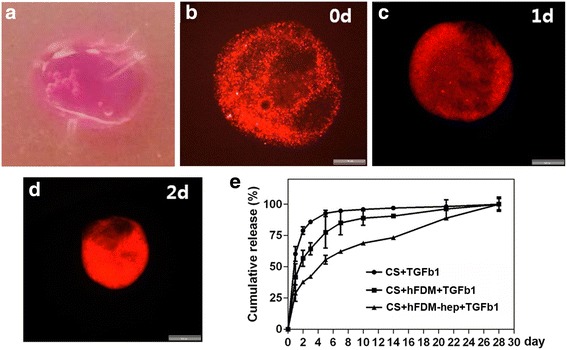
Fig. 3.

Preparation of collagen spheroids. a Collagen spheroids containing hPMSCs and hFDM-hep; b–d Collagen spheroids containing hPMSCs, hFDM-hep and live cell tracking dye (PKH26) at different time points in vitro; e Release profile of TGF-β1 from three different groups of collagen spheroids for 28 days in vitro. CS: collagen spheroid. Scale bar is 200 μm
