Fig. 5.

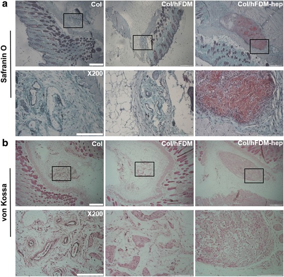
Histological analysis of subcutaneously implanted collagen spheroids at 4 weeks. a Safranin-O staining and b von Kossa staining. The boxed area appears in higher magnification. Scale bar is 500 and 200 μm, respectively

Histological analysis of subcutaneously implanted collagen spheroids at 4 weeks. a Safranin-O staining and b von Kossa staining. The boxed area appears in higher magnification. Scale bar is 500 and 200 μm, respectively