
Figure 2. Overview of exosome formation.
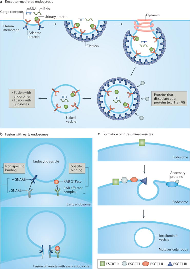
Exosome formation occurs through a multistep process that is initiated by pinocytosis (not shown) or receptor-mediated endocytosis (part a), which involves the binding of urinary proteins to the apical membrane and their internalization, a process that requires the coat protein clathrin and the ATPase dynamin. Invagination of the lipid bilayer results in formation of a small unilamellar vesicle. Proteins such as heat shock protein 70 (HSP70) can dissociate coat proteins (for example, clathrin) to yield a naked vesicle that can fuse with early endosomes (part b), a process that is mediated by soluble N-ethylmaleimide-sensitive factor attachment protein receptors (SNAREs) and small RAB effector proteins. Intraluminal vesicles form after invagination of the endosomal membrane (part c), a process that is carried out by tetraspanins (not shown) and endosomal sorting complex required for transport (ESCRT) protein complexes, such as ESCRT-0 (which is responsible for cargo clustering), ESCRT-I and ESCRT-II (both are responsible for inducing bud formation), and ESCRT-III (which promotes intraluminal budding of vesicles in endosomes and vesicle scission)171. The dissociation and recycling of the ESCRT machinery is carried out by accessory proteins. miRNA, microRNA; t-SNARE, target SNARE; v-SNARE, vesicle SNARE.