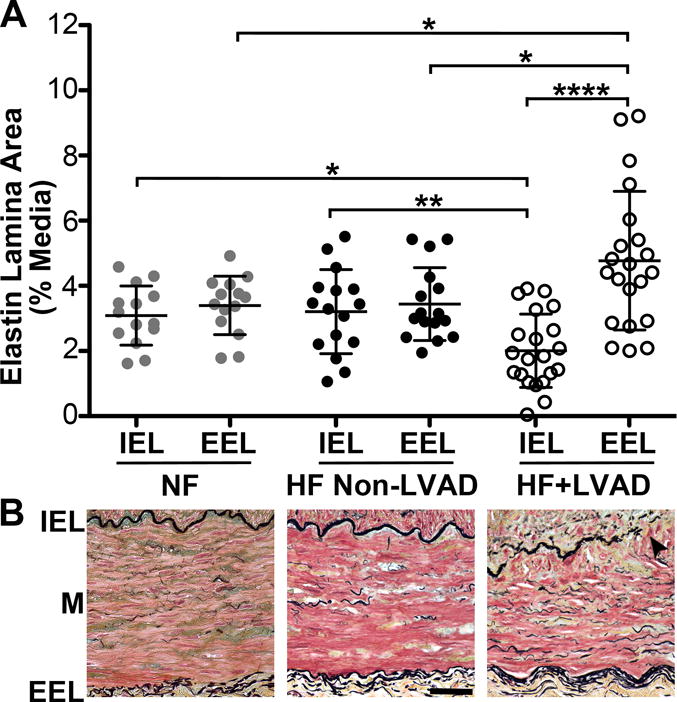
Figure 2. Medial Elastic Lamina Remodeling.

A) The areas of the internal elastic lamina (IEL) and external elastic lamina (EEL) are reported as the percent-area relative to the media area of the transverse coronary artery section. There were significant differences in the areas of both the IEL and EEL between the Non-Failing, HF Non-LVAD, and HF+LVAD groups. In the HF+LVAD group, there was significant attenuation of the IEL and thickening of the EEL. B) Representative high power 40x images of coronary arteries from Non-Failing, HF Non-LVAD, and HF+LVAD patients. The IEL containing black-stained fibers run at the top of the image along the intima-media (M) border and the lower EEL run along the media-adventitia border. Arrowheads show breakage and attenuation of the IEL compared to the thickening of the EEL in HF+LVAD vessels.
