
Figure 3. Changes in Collagen Deposition within the Adventitia of the Coronary Arteries.
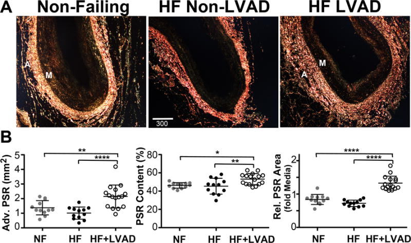
A) Representative polarized darkfield images of PicroSirius Red (PSR) stained coronary artery sections from Non-Failing, HF Non-LVAD, and HF+LVAD individuals demonstrate differences in collagen deposition as measured by PSR birefringence within the adventitia (A). Images are shown at 4x magnification. Scale bar represents 300 μ. B) There was a significant increase in adventitial collagen content among HF+LVAD patients compared to Non-Failing and HF Non-LVAD patients as assessed by the area of PSR birefringence in the adventitia (left panel) and the percent content of PSR birefringence (positive fractional area of PSR signal/adventitia area, middle panel). These differences among groups remained significant when controlling for differences in vessel size by measuring the relative area of PSR birefringence in the adventitia normalized to the vessel media (M) area (right panel).