
Figure 4. Changes in Coronary Artery Adventitia Fibrillar Collagen as Detected by Second Harmonic Generation Imaging.
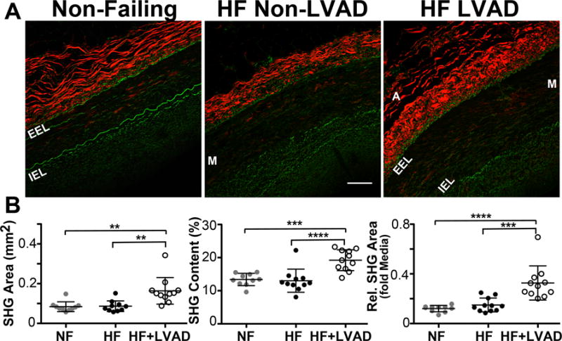
A) Representative second harmonic generation (SHG) images of coronary artery sections from Non-Failing, HF Non-LVAD, and HF+LVAD individuals. The fibrillar collagen within the adventitia (A) produces an autofluorescent SHG signal that is displayed as red in color. The internal elastic lamina (IEL) and external elastic lamina (EEL) surrounding the vessel media (M) is displayed as green in color. Images shown at 20x magnification. Scale bar represents 100 μ. B) The area of SHG+ collagen in the coronary adventitia of HF+LVAD patients is increased relative to Non-Failing and HF Non-LVAD patients (left panel). Furthermore, the percent content of SHG+ collagen in the adventitia, a correlate of collagen density, is higher in the HF+LVAD group compared to Non-Failing and HF Non-LVAD (middle panel). These differences remained significant when controlling for differences in vessel size by measuring the relative area of adventitial SHG+ collagen normalized to the media area (right panel).