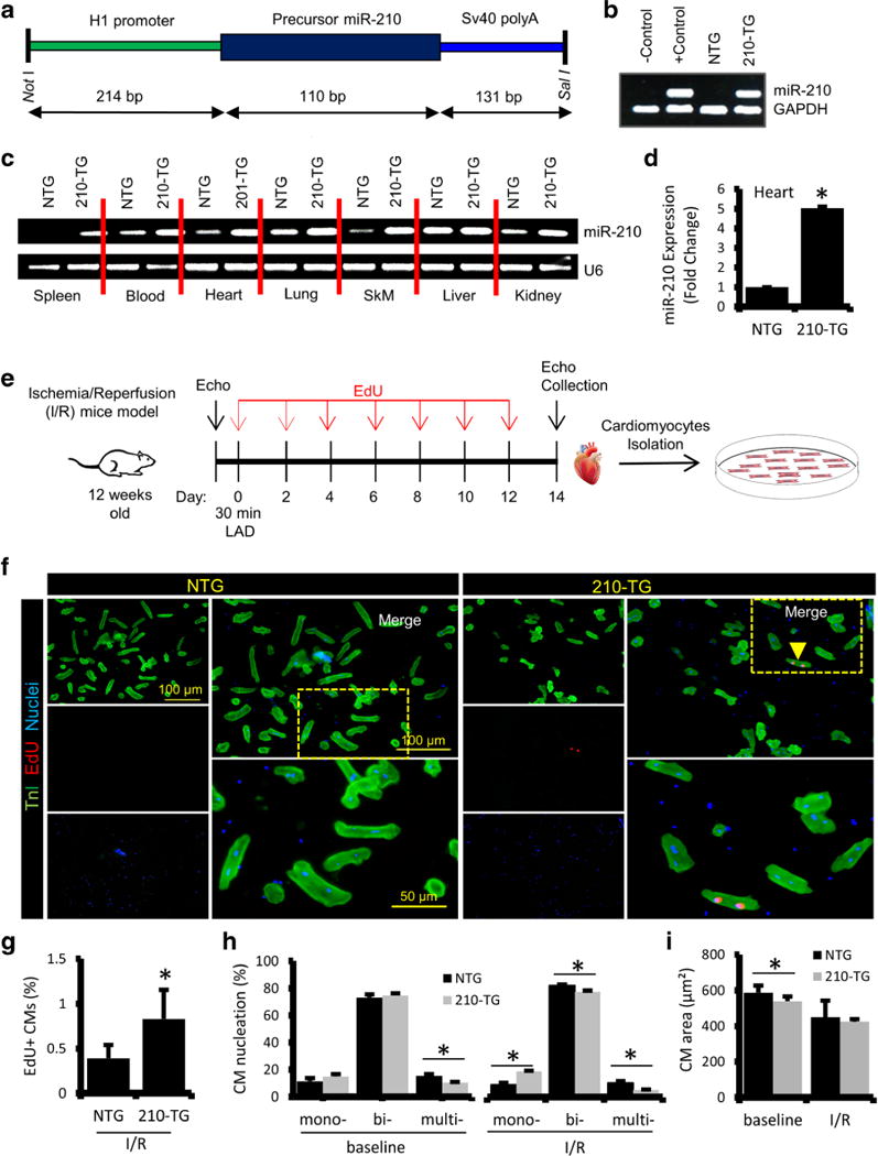
Fig. 4.

MiR-210 overexpression promotes CM proliferation in I/R injury mice model. a MiR-210 overexpressing transgenic (210-TG) mice were generated using C57BL/6 strain. 210-TG construct was designed to have an H1 promoter, precursor miR-210 cDNA, and SV40 poly A cassette at 3′ end. Precursor miR-210 along with H1 promoter was amplified and cloned into pShuttle vector using restriction enzymes Not1 and Sal1. Amplified vector was digested, purified, and used for microinjection into the pronuclei of single-cell embryos derived from superovulated C57BL/6 females. The surviving embryos were implanted into foster female mice. b Genotyping of mice tail genomic DNA via PCR using custom designed primers for miR-210, showing miR-210 transgenic (210-TG; lane 4) and non-transgenic (NTG; lane 3). GAPDH was used as internal control. c qPCR results showing miR-210 expression in different organs such as the spleen, heart, lung, skeletal muscle (SkM), liver, kidney, and blood of 210-TG and NTG mice. d Quantification ofmiR-210 expression by qPCR showing miR-210 overexpression in 210-TG mice hearts. e Schematic representation of 12-week-old mice subjected to LAD ligation for 30 min followed by reperfusion for 2 weeks (ischemia/reperfusion injury model). EdU (350 μg/100 μl PBS; i.p.) treatments were given at every alternate day till day 12. Post 2 weeks of surgery, echocardiography was performed, and the hearts were collected for CM isolation and culture. f Immunostaining of CMs, against TnI (green) and EdU (Red; indicated by arrowhead). Cell nuclei were counterstained with DAPI (blue). g–i Quantification of EdU labeling, cell nucleation (Mono-, bi-, multi-), and CM size, showing a significant increase in EdU+ CMs and mono-nucleation, while multi-nucleation was decreased in 210-TG hearts following I/R injury. N = 4 for all studies. *P < 0.05 vs NTG
