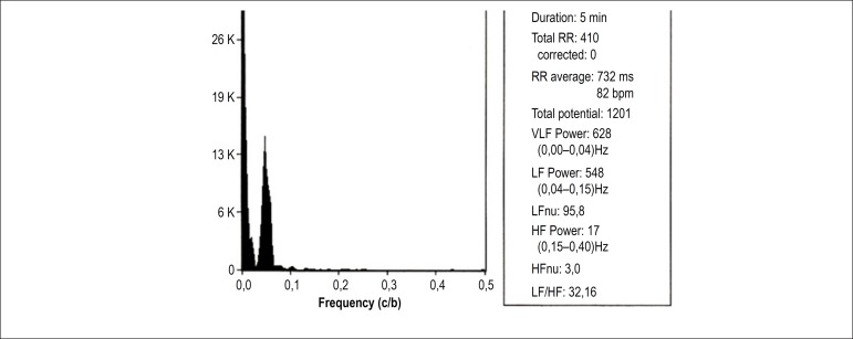
Figure 4.
Spectral analysis of a female patient (62 years of age) without orthostatic hypotension (the same of Figure 3) in orthostatic position, showing an increase in low frequency (LF) component and in the LF/high frequency (HF) ratio, in relation to supine position. VLF: very low frequency; RR: number of QRS in sinus rhythm; VLF: very low frequency; HFnu: HF normalized unit.

