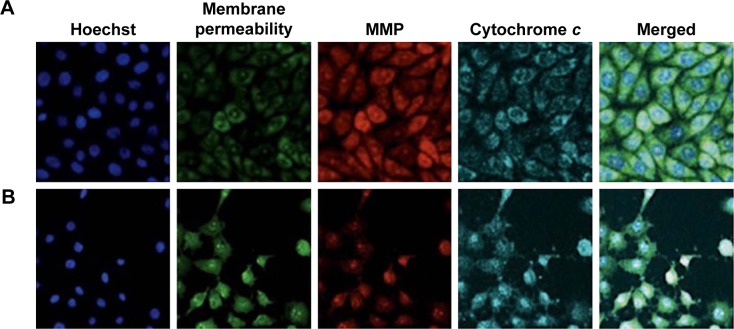
Figure 9.
Representative images of PC-3 cells treated with medium alone (A) and PC-3 cells induced using koenimbin (B).
Note: The PC-3 cells are stained with Hoechst for nuclei, permeability membrane, MMP, cytochrome c and merged images (magnification, ×20).
Abbreviation: MMP, mitochondrial membrane potential.

