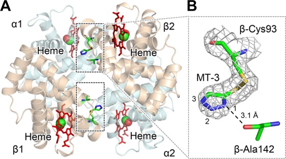
Figure 4.

Crystal structure of COHbA in a complex with TD-3. MT-3 formed a disulfide bond with the thiol of COHbA β-Cys93. (A) The binding sites of MT-3 in COHbA. Hb α and β subunits are shown as pale blue and brown, respectively. Carbon monoxide bound to heme is shown as a red and green sphere. The locations of MT-3, β-Cys93, and β-Ala142 are shown as sticks within the dashed rectangles. (B) The electron density of MT-3 and β-Cys93 indicated by the 2Fo-Fc map (gray mesh, contoured at 1.0σ). The N1 atom of MT-3 at β-Cys93 formed a hydrogen bond with the oxygen atom of β-Ala142, which is shown as a dotted line.
