Figure 6.
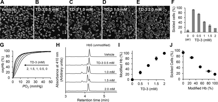
Inhibition of hypoxia-induced sickling of SS RBCs by TD-3 in vitro. The morphology of SS RBCs treated with vehicle (A) or with TD-3 (0.5–2 mM, B–E) and incubated with 4% oxygen at 37 °C for 2 h. (F) The effect of TD-3 on the percentage of sickled cells (sickled cells%) in cells exposed to hypoxia. (G) Representative oxygen dissociation curves (ODCs) of the hemolysate of SS RBCs with or without TD-3. The ODCs were measured in phosphate buffer (0.1 M phosphate, pH 7.0) at 25 °C. (H) HPLC chromatograms of hemolysates prepared from SS RBCs treated with TD-3 (0–2 mM). (I) The effect of treating SS RBCs with TD-3 on the percentage of modified Hb (modified Hb%). (J) The relationship between the modified Hb% and sickled cell% of SS RBCs treated with TD-3. Each data point represents the mean value of modified Hb% or sickled cells% measured in triplicate. Error bars represent standard deviation. Panel J was generated using the data from panels F and I.

