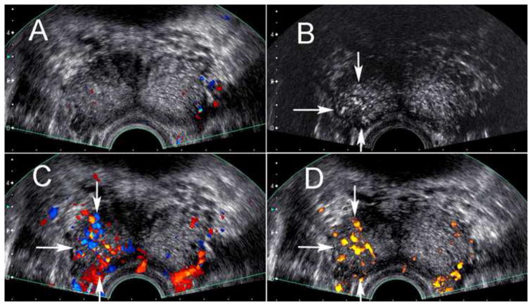
Figure 2.
Transrectal ultrasound near the base of the prostate in a 62 year old male. Targeted and systematic biopsy cores at the right base demonstrated greater than 50% core involvement with Gleason 8 and 9 prostate cancer. The pre-contrast grayscale and color Doppler image (A) is unremarkable, but the post-contrast harmonic grayscale image (B) as well as the post-contrast color (C) and power (D) Doppler images demonstrate hypervascularity associated with the area of positive targeted cores (arrows).

