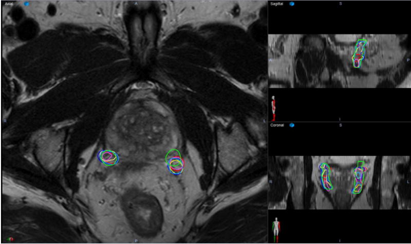
Figure 2.

A representative axial (left), sagittal (upper right), and coronal (lower right) slices for a 3-Tesla pelvic magnetic resonance imaging scans with overlaying contours of the left and right neurovascular bundle from each rater and the expert. On this image, the red contours on each side are the expert. Dark blue represents rater 1, yellow represents rater 2, light blue represents rater 3, pink represents rater 4, and lime green represents rater 5.
