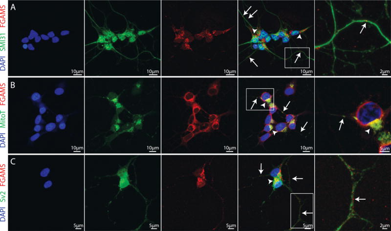
Figure 1. FGAMS protein expression occurs in neuronal processes and near mitochondria in differentiated SH-SY5Y human neuroblastoma cells.

(A) Differentiated SH-SY5Y neurons immunolabeled for the axonal marker SMI31 (green) and the purine biosynthetic enzyme FGAMS (red). FGAMS demonstrated strong localization in the neuronal cytoplasm (arrowheads) with numerous punctate localizations throughout neuronal axons (arrows). (B) Consistent with recent work in primary hippocampal neurons (Williamson et al. 2017), FGAMS (red) co-localized with mitochondria (green), both in neuronal cytoplasm (arrowheads) and along neurites (arrows) in differentiated SH-SY5Y neurons. (C) When neurons were immunolabeled for both FGAMS (red) and Sv2 (synaptic vesicle protein 2 – green), no distinct enrichment of FGAMS at synapses was evident. All cells were counterstained with DAPI nuclear stain (blue). Two coverslips from 3-4 independent cultures were analyzed.
