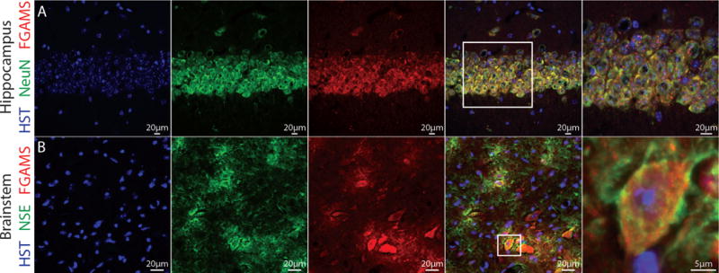
Figure 3. In vivo expression of FGAMS is evident in neuronal cell bodies.

To assess FGAMS expression in neurons in vivo, whole brain sections derived from 3 month old male mice were probed for FGAMS (red) and two neuronal markers (green). (A) Images acquired from the CA1 region of the hippocampus demonstrated co-localization of FGAMS and neuronal nuclei (NeuN) in the cell body layers. (B) In the medulla region of the brainstem, co-localization of neuron-specific enolase (NSE) and FGAMS was observed. All sections were counterstained with Hoescht to mark the nucleus (blue). Sections were acquired from two different animals, and 2-4 sections per animal were processed for analysis.
