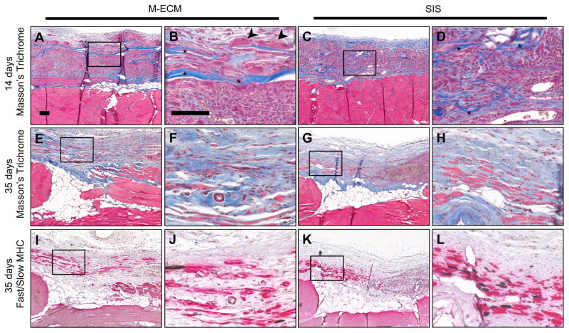
Fig. 8.
In vivo response to implanted (A–B,E–F,I–J) M-ECM and (C–D,G–H,K–L) SIS after (A–D) 14 and (E–L) 35 days. Sections stained with (A–H) Masson’s Trichrome or (I–L) immunolabeled for fast (red) and slow (brown) myosin heavy chain (MHC). There are obvious scaffold remnants at 14 days for both scaffolds (asterisks), and multinucleate cells around M-ECM scaffold regions (arrowheads). Scale bar represents 100 μm. (For interpretation of the references to color in this figure legend, the reader is referred to the web version of this article.)

