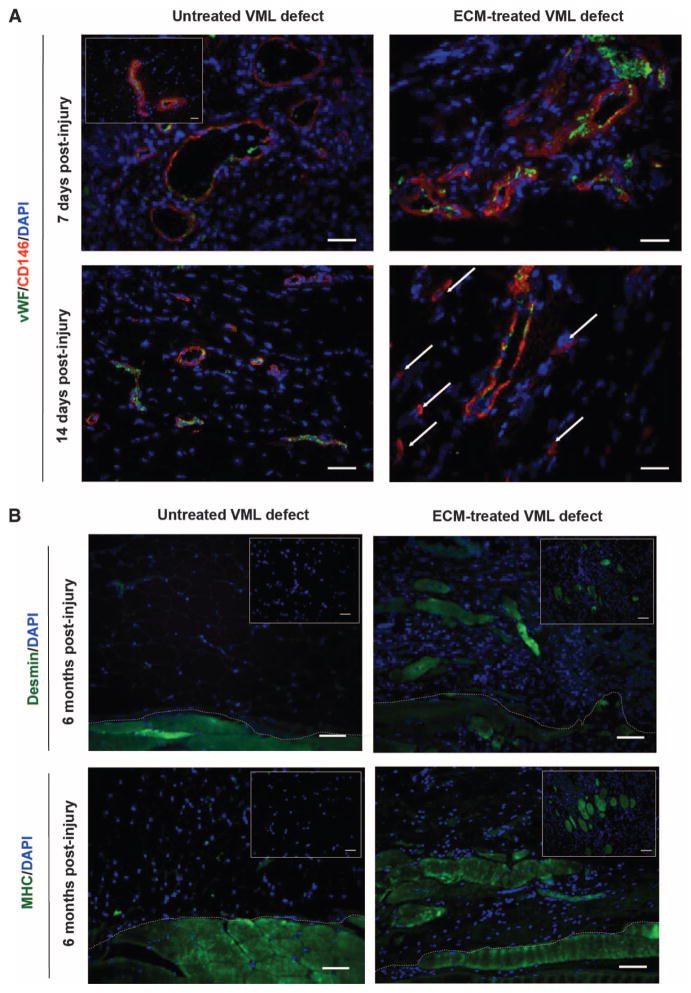
Fig. 1. Progenitor cells are present at the site of ECM scaffolds surgically placed within sites of mouse VML injury.
(A) CD146+ PVSCs remained in their normal anatomic location, surrounding vascular structures (identified by vWF) at 7 days after injury in both untreated and ECM-treated defects in a mouse model of VML. Healthy, normal mouse skeletal tissue is shown in inset. After 14 days, PVSCs maintained their vascular association in untreated VML defects; conversely, in ECM-treated defects, CD146+ PVSCs were present outside their normal niche (arrows). (B) Untreated defects from the mouse model of VML showed no signs of skeletal muscle formation (desmin+, MHC+ cells), whereas ECM scaffold–treated defects showed desmin+, MHC+, and striated skeletal muscle cells at 6 months after injury. Skeletal muscle cells were identified along the interface with underlying native tissue (dotted white line) as well as throughout the center of the ECM implantation site (insets). Scale bars, 50 μm. DAPI, 4′,6-diamidino-2-phenylindole; MHC, major histocompatibility complex.

