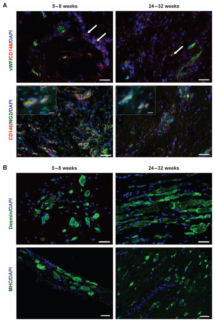
Fig. 4. Constructive tissue remodeling by ECM scaffolds in patients.
Representative images are shown here, with images from each patient in fig. S2. (A) Biopsied human muscle tissue from ECM-treated VML defects at both 5 to 8 weeks and 24 to 32 weeks after scaffold implantation. PVSCs (CD146+NG2+) were present both within and outside of (arrows) their normal perivascular association (vWF+ regions). High-magnification insets show colocalization of CD146 and NG2 (inset scale bars, 10 μm). Scale bars, 50 μm. (B) Human muscle biopsies from the site of scaffold implantation at 5 to 8 weeks and 24 to 32 weeks after ECM scaffold implantation showed the formation of islands of desmin+ muscle cells. Scale bars, 50 μm.

