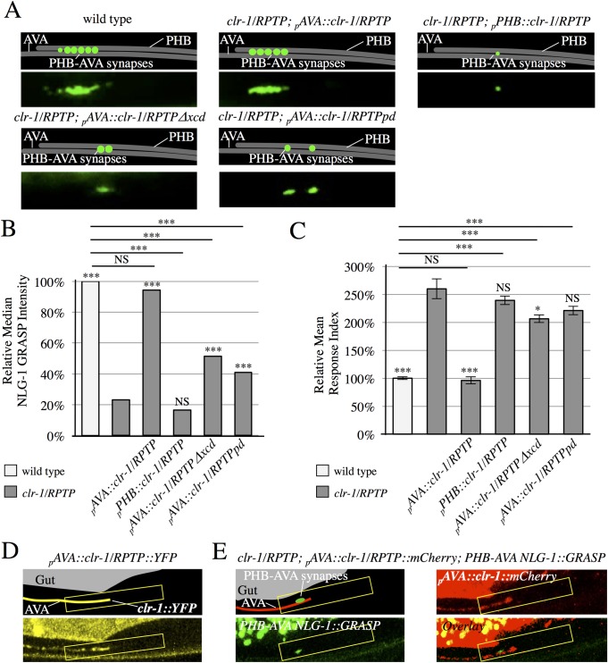
Fig 2. CLR-1/RPTP acts in postsynaptic neurons, and is localized to the synaptic region.
(A) Schematics and micrographs of normal PHB-AVA NLG-1 GRASP fluorescence in wild-type and clr-1/RPTP(e1745) mutant animals expressing a transgene that drives expression of the clr-1 cDNA in AVA neurons (pAVA::clr-1/RPTP), and reduced PHB-AVA NLG-1 GRASP fluorescence in clr-1/RPTP(e1745) mutant animals expressing either a construct that drives expression of the clr-1/RPTP cDNA in PHB neurons (pPHB::clr-1/RPTP), a transgene that drives expression of the clr-1/RPTP cDNA with the extracellular domain deleted in AVA neurons (pAVA::clr-1/RPTPΔxcd), or a transgene that drives expression of the clr-1/RPTP cDNA with a mutation that inactivates the phosphatase domain (pAVA::clr-1/RPTPpd). (B) Quantification of NLG-1 GRASP fluorescence. Expression of clr-1/RPTP in AVAs, but not PHBs restores NLG-1 GRASP fluorescence in clr-1/RPTP(e1745) mutants (n>75). Expression of the clr-1/RPTP cDNA with the extracellular domain deleted or with a mutation in the active site of the phosphatase domain does not fully restore NLG-1 GRASP fluorescence in clr-1/RPTP(e1745) mutants (n>100). Two or more lines were examined with each transgene, and combined in the graph above. Values for each individual transgenic line are included in S2 Table. NS, not significant, ***P<0.001, *P<0.05, U-test. Comparison to clr-1/RPTP indicated over individual bars. P-values were adjusted for multiple comparisons using the Hochberg method. 95% confidence intervals for the medians are included in S1 Table. (C) Expression of clr-1/RPTP in AVAs, but not PHBs, rescues the behavioral defect in clr-1/RPTP(e1745) mutants (n>75). Expression of clr-1/RPTP cDNA with the extracellular domain deleted or with a mutation in the active site of the phosphatase domain does not fully rescue the behavioral defect in clr-1/RPTP(e1745) mutants (n≥60). NS, not significant, ***P<0.001, t-test. Comparison to clr-1/RPTP indicated over individual bars. P-values were adjusted for multiple comparisons using the Hochberg method. Error bars are SEM. (D) Schematic and micrograph of an animal expressing the clr-1/RPTP cDNA linked to YFP in AVA (pAVA::clr-1/RPTP::YFP). (E) Schematic and micrograph of an animal expressing the clr-1/RPTP cDNA linked to mCherry in AVA (pAVA::clr-1/RPTP::mCherry) and PHB-AVA NLG-1 GRASP, and overlay in a clr-1/RPTP mutant animal. (D-E), CLR-1 localization is brightest in the preanal ganglion (yellow box), and the majority of animals show localization in the anterior half of this region, where PHB-AVA synapses usually form (green fluorescence).

