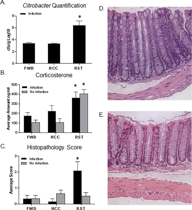
Fig 7. Stressor exposure increases C. rodentium levels and colonic histopathology scores.
A. Citrobacter levels significantly increased in feces of stressor exposed mice (*p<0.05, vs HCC Infection and FWD Infection). B. Corticosterone levels were significantly increased in mice exposed to the RST stressor (*p<0.05, main effect of stress). C. Mice exposed to the infection and exposed to the RST stressor had a significant increase in histopathology scores (*p<0.05, vs HCC No Infection, FWD No Infection, RST No Infection, HCC Infection, and FWD Infection). D. A representative image of the histopathology for a mouse with mild disease exposed to the infection and RST stressor. E. A representative image of the histopathology for a mouse with no inflammation. All representative histologic sections are from the distal colon and are shown at the same magnification of 20X. Data are the mean +/- SEM.

