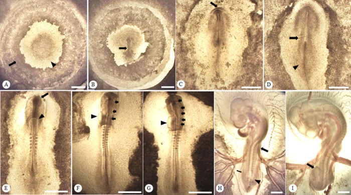
Fig 2. Developmental structures and rudiments of embryonic features during the first 72 h of incubation.

Representative figures show the structures of Taiwan Country Chicken (TCC) embryos. (A) The area opaca (arrow) and the area pellucida (arrowhead) are distinct at 5 h post-incubation; (B) the primitive streak (arrow) appears at 12 h in TCC post-incubation; (C) the headfold (arrow) becomes visible at 25 h post-incubation; (D) the initial pair of somites (arrow) and neural plate (arrowhead) first appear at 26 h post-incubation; (E) the primary optic vesicles (arrow) and the paired heart primordia (arrowhead) start to form at 30 h post-incubation; (F) the three primary brain rudiments (arrows) are visible at 33 h post-incubation along with the developing heart primordia (arrowhead) at the 13-somite stage; (G) the five neuromeres (arrows) are distinguishable at 42 h post-incubation with a relatively developed heart (arrowhead); (H) the wing (arrow) and leg buds (arrowhead) develop at 57 h post-incubation and the allantois is barely visible (thin arrow); (I) a prominently enlarged allantois (arrow) can be identified at 72 h post-incubation. Bright field, scale bar = 0.5 mm.
