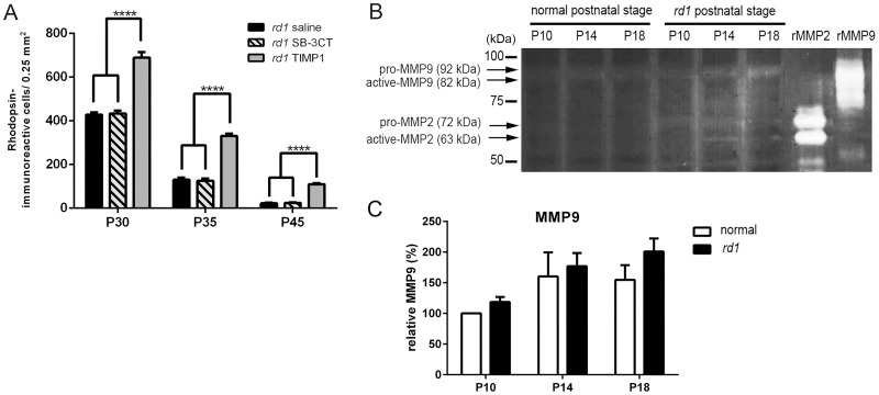
Fig 1. SB-3CT-treatment did not affect the rod survival in the rd1 mouse retina.
The summary graphs illustrate mean rod density measured from the 0.25 mm2 sampling areas (in the superior-temporal region) of all saline-treated, SB-3CT-treated, and TIMP1–treated rd1 retina groups (A). Retinal extracts of normal and rd1 were collected at P10, P14, and P18 for gelatin zymography (B). Recombinant mouse MMP9 and recombinant mouse/rat MMP2 were applied to the gel and transferred to the membrane as positive controls. Data represent mean ± SEM, ****P<0.0001; P, postnatal.

