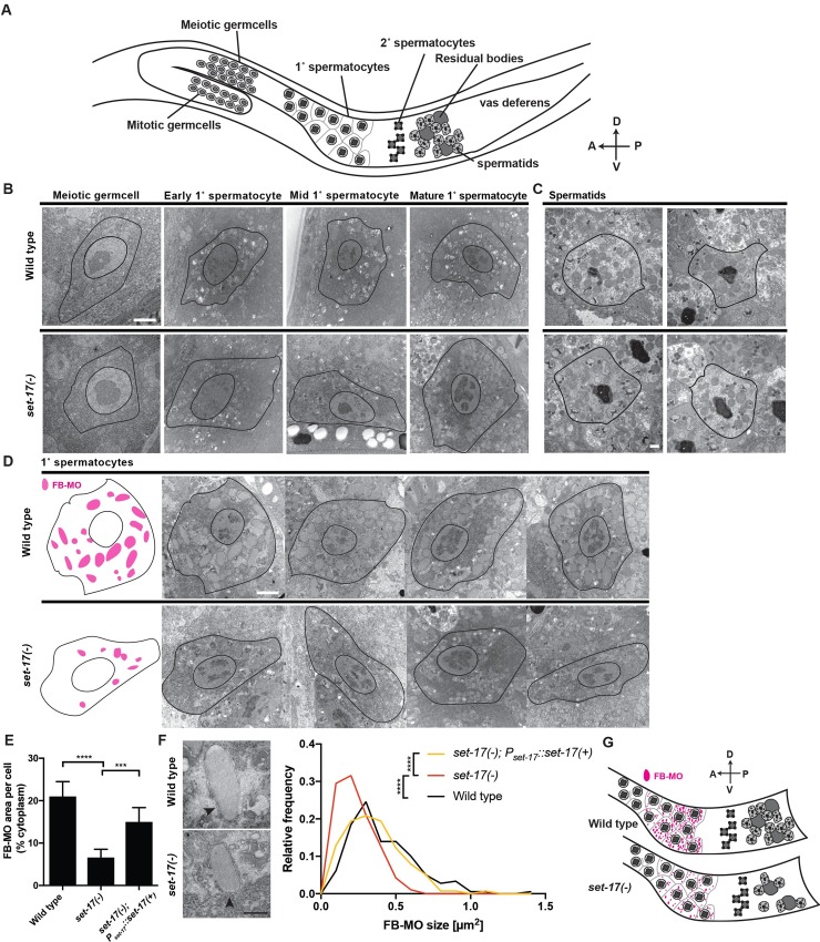
Fig 4. set-17 promotes the number and size of major sperm protein vesicles in primary spermatocytes.
A) Schematic of the male germline. Meiotic germ cells differentiate into primary spermatocytes, which mature as they progress along the gonad into secondary spermatocytes and then spermatids. A, anterior; P, posterior; D, dorsal; V, ventral. B) Electron micrographs of spermatocyte differentiation in wild-type and set-17 males. All spermatocyte differentiation stages are present in set-17 mutants. Images were staged based on their relative positions along the germline. Scale bar, 2 μm. C) Electron micrographs of spermatids in wild-type and set-17 males. Spermatids appear normal in set-17 mutants. Scale bar, 500 nm. D) Electron micrographs of mature primary spermatocytes from wild-type and set-17 males. Left, schematic traces of the outlines of the left-most panels of wild-type and set-17 primary spermatocytes (black), respectively, indicating the areas and positions of FB-MOs (magenta). Fibrous-body membranous organelles (FB-MOs) are smaller in set-17 mutants. Scale bar, 2 μm. E) Percent cytoplasmic cross-sectional area taken up by FB-MOs per cell in wild-type, set-17 and set-17; Pset-17::set-17(+) male mature primary spermatocytes. n = 10; **** P < 0.0001, *** P < 0.001, t-test. F) Frequency distributions of FB-MO cross-sectional areas (size) in wild-type, set-17 and set-17; Pset-17::set-17(+) male primary spermatocytes. Arrowheads, FB-MOs in micrographs of representative wild-type and set-17 spermatocytes. n > 95; **** P < 0.0001, KS-test; Mean: wild-type = 0.41 μm2, set-17 = 0.24 μm2, set-17; Pset-17::set-17(+) = 0.35 μm2; Scale bar, 500 nm. G) Schematic of sperm production in wild-type and set-17 males. In set-17 mutants, defective FB-MO production in primary spermatocytes leads to smaller FB-MOs and a reduction in the production of otherwise normal spermatids. A, anterior; P, posterior; D, dorsal; V, ventral.

