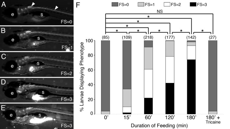
Figure 1.

A qualitative food intake assay for zebrafish larvae. (A-E) Side views of 7 dpf larvae examined under GFP epifluorescence. The larvae were incubated with Y-G fluorescent microspheres coated with fish food (see Methods) for up to 3 hours before visualizing the fluorescent contents in their gut. The amount of fluorescence (ignoring autofluorescence) ranged from little or none (assigned a feeding score (FS) of 0), less than 25% of the gut (FS=1), about 50% of the gut (FS=2), to a full gut (FS=3). Most experiments were performed with Tg(isl1:gfp) larvae that express GFP in cranial and spinal motor neurons (arrowheads) and enabled proper sample orientation prior to scoring. e, eye; s, swim bladder. (F) Pooled data from 2-8 experiments (number of larvae in parenthesis) showing the distribution of feeding scores for various durations of feeding. For tricaine treatment, larvae were exposed to the chemical in the medium for 5 minutes before addition of labeled food. The observers assigning feeding scores were blinded to the treatment conditions of the larvae being scored. As the duration of feeding increased to 3 hours, the fraction of larvae exhibiting feeding scores of 2 and 3 increased. As expected, Tricaine-treated larvae, which were paralyzed and unable to swim, ate very poorly. Asterisk, Chi-square test with Bonferroni correction indicating significance at p < 0.003; NS=not significant.
