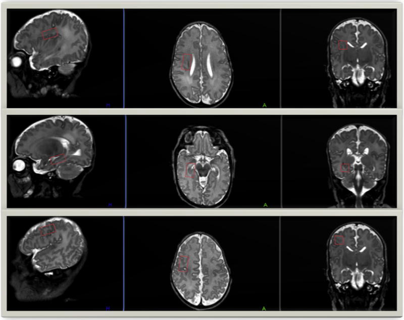
FIGURE 1.

Single-voxel placement locations in the three cerebral regions of interest. T2-weighted MRI images in the sagittal, axial, and coronal planes display the single-voxel placements in our three regions of interest—the right subventricular zone (top), the right hippocampus (center), and the right frontal cortex (bottom). MRI, magnetic resonance imaging. (Color version of the figure is available in the online edition.)
