
Figure 1.
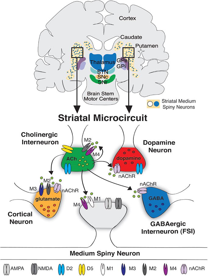
Cholinergic Interneurons in the Striatal Microcircuit. Top: Coronal schematic of the human basal ganglia. Striatal medium spiny neurons are highlighted in the caudate and putamen as blue and white circles. Bottom: Illustration of the distribution of striatal cholinergic receptors and sites of cholinergic regulation (arrows). Abbreviations: D2 (dopamine type 2-like receptor), D5 (dopamine type 5-like receptor), nAChR (nicotinic acetylcholine receptor), M1,2,3, or 4 (muscarinic acetylcholine receptor), ACh (acetylcholine), FSI (fast spiking interneuron) NMDA (N-methyl-D-aspartate receptor), AMPA (α-amino-3-hydroxy-5-methyl-4-isoxazolepropionic acid receptor), GABA (γ-Aminobutyric acid).