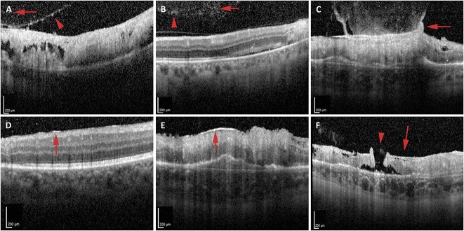
Fig. 1.
Vitreoretinal interface changes in CMV retinitis. A and B. Optical coherence tomography identified abnormalities within the posterior vitreous including hyperreflective deposits on the posterior surface of the hyaloid (A, red arrowhead), posterior vitreous cells (A and B, red arrows), and clumps of hyperreflective material consistent with vitreous debris (B, red arrowhead). C–F. Vitreoretinal interface changes noted on OCT include gliosis of the posterior hyaloid with taut, broad-based vitreomacular traction to the underlying area of retinitis (C, red arrow), ERM over retina unaffected (D, red arrow) or affected (E, red arrow) by retinitis, and a lamellar hole–associated epiretinal proliferation (F, red arrow) associated with a full thickness atrophic eccentric macular hole (F, red arrowhead) in an area of atrophy from retinitis.

