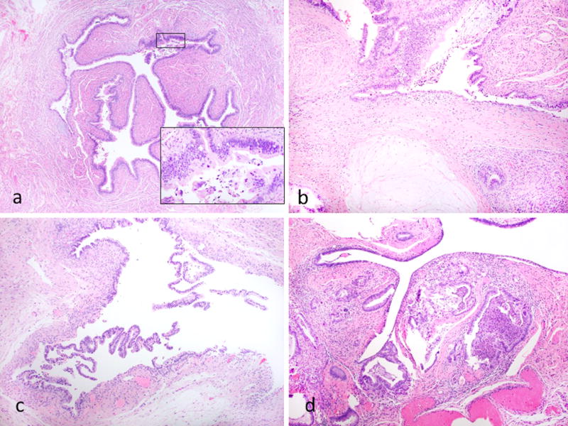
Figure 3.

Other features of fallopian tube involvement. Case showing focal involvement of the non-fimbrial portion of the tube; there is epithelial proliferation with nuclear atypia and tufting (inset) (a). Case showing mucin extravasation within the tubal stroma (b). Case where the mucinous epithelium has lifted-off” and separated from the underlying stroma resulting in a subepithelial cleft (c). Case exhibiting involvement of submucosa (d).
