
Fig. 1. Laser doppler imaging of blood flow in a patch test.
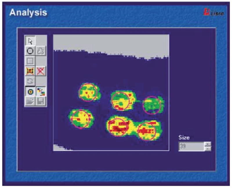
Printed with permission from Fullerton et al. (2002) (10). The perfusion image can be analysed by an integrated system software. The relative colour scale extends from the smallest and the largest perfusion value (from green to red). (https://www.perimed-instruments.com/skin-patch-testing).