
Fig. 2. Laser Speckle Imaging (LSI) of a Caucasian female patient with a port wine stain involving the V2 dermatomal distribution.
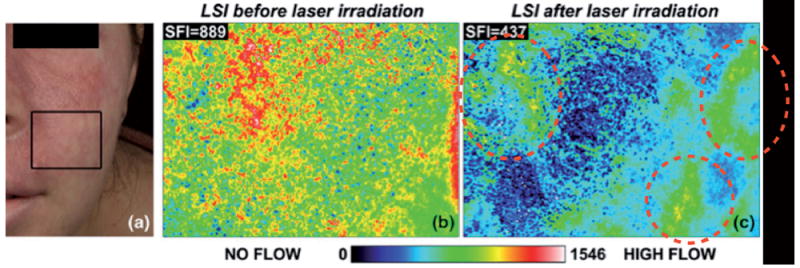
Printed with permission from Huang Y.C. et al. (2008) (11). (a) Photograph. (b) Speckle Flow Index images taken from the marked region of interest immediately before and (c) 15 min after laser therapy. Colour range indicates the level of blood flow in this area.