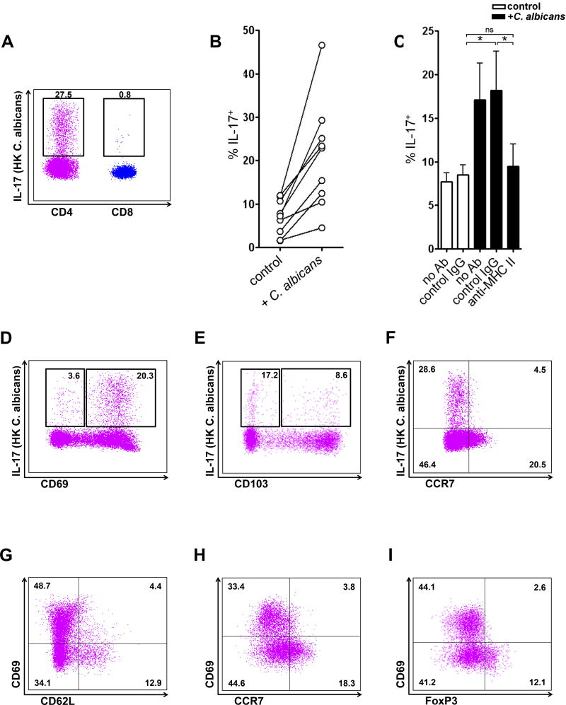
Figure 7. C. albicans-specific Th17 cells are abundant in normal human skin.
(A) Expression of IL-17 in CD4 and CD8 T cells (gated on CD3+CD4+ and CD3+CD8+ cells) from normal human skin when treated with HK C. albicans. Data are representative of five independent experiments. (B) Expression of IL-17 in CD4 T cells from normal human skin when cultured in the presence (+ C. albicans) or absence (control) of HK C. albicans. (C) C. albicans mediated increased IL-17 production was dependent on MHC class II. Neutralizing antibodies to MHC II (anti-MHC II) blocked increases in IL-17 production in C. albicans-treated cultures (black bars). The mean and SEM of 5 donors are shown. *p < 0.05; ns, not significant. (D) Expression of IL-17 in CD69+ and CD69− CD4 T cells (gated on CD3+CD4+ cells) from normal human skin. (E) Expression of IL-17 in CD103+ and CD103− CD4 T cells (gated on CD3+CD4+ cells) from normal human skin. (F) Expression of IL-17 in CCR7+ and CCR7− CD4 T cells (gated on CD3+CD4+ cells) from normal human skin. (G) Expression of CD62L in CD69+ and CD69− CD4 T cells (gated on CD3+CD4+ cells) from normal human skin. (H) Expression of CCR7 in CD69+ and CD69− CD4 T cells (gated on CD3+CD4+ cells) from normal human skin. (I) Expression of FoxP3 in CD69+ and CD69− CD4 T cells (gated on CD3+CD4+ cells) from normal human skin. Data are representative of three independent experiments (D–I). * p < 0.05; ns, not significant.

