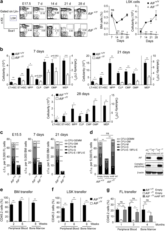
Fig. 3. Mitochondrial OXPHOS dysfunction associated with AIF loss generated cell exhaustion and alteration in the repopulating potential of hematopoietic progenitors.
a Left, Identification of LSK (Lin−Sca-1+ Kit+) cells in fetal liver (E17.5) or BM from 7-, 14-, 21-, and 28-day-old AIF+/Y and AIF−/Y animals by flow cytometry. Numbers are the percentages of LSK cells. Right, The number of LSK cells was recorded and graphed either as a percentage of total cells in the BM or as a number of cells (cellularity) (n = 5 mice per group). b Number of LT-HSC, ST-HSC, MPP, CLP, CMP, GMP, and MEP cells in 7- to 28-day-old animals (n = 5 mice per group). c Fetal liver cells (FL; E15.5) or BM cells from 7- or 21-day-old animals were seeded in methylcellulose culture and colonies formed by the GEMM, GM, or single lineages (G, M, and E) were counted (n = 8 independent experiments). d The effects of AIF re-expression on the colony-forming capacities were tested with E15.5 FL cells from AIF+/Y and AIF−/Y embryos transduced with the retroviral MSCV-IRES-GFP (empty) or MSCV-mAIFWT-IRES-GFP (mAIF WT) vectors. Colonies were counted as in c (n = 3 independent experiments). The expression of AIF and the complex I subunit NDUFA9 was verified by immunoblotting. Equal loading was confirmed by β-actin probing. e In non-competitive transplantation experiments, CD45.2 donor BM cells from 7-day-old AIF+/Y and AIF−/Y mice were transplanted into irradiated CD45.1 recipient animals. CD45.2 chimerism in recipient mice was evaluated at the indicated time after transplantation (n = 5 mice per group). f In competitive transplantation experiments, CD45.2 donor LSK cells from 7-day-old WT or AIF KO animals were transplanted into irradiated CD45.1 mice along with recipient BM cells. CD45.2 chimerism in recipient mice was assessed at 2, 6, and 8 weeks after transplantation (n = 5 mice per group). g The effects of mAIF re-expression on hematopoietic reconstitution were tested with E15.5 FL cells from AIF+/Y and AIF−/Y embryos transduced with the retroviral MSCV-IRES-GFP (Empty) or MSCV-mAIFWT-IRES-GFP (mAIF WT) vectors (n = 6 mice per group). Statistical significance was calculated by Mann–Whitney a, b, c, e, f, and g or two-way ANOVA d tests. Symbols and bars represent mean ± SEM

